Read online Transfontanellar Doppler Imaging in Neonates (Medical Radiology) - Alain Couture file in PDF
Related searches:
Transfontanellar doppler imaging in neonates medical radiology dejavusansextralight font size 10 format.
Doppler tissue imaging: myocardial wall motion velocities in normal subjects. Color-coded measures of myocardial velocity throughout the cardiac cycle by tissue doppler imaging to quantify regional left ventricular function.
The doppler evaluation of the intracranial arteries is performed with a low-frequency (2–3 mhz) sector imaging transducer. The measured flow velocities are only correct if there is a zero degree angle between the vessel and the doppler beam.
A doppler ultrasound is a noninvasive test that can be used to estimate the blood flow through your blood vessels by bouncing high-frequency sound waves (ultrasound) off circulating red blood cells. A regular ultrasound uses sound waves to produce images, but can't show blood flow.
Tissue doppler (tissue velocity imaging) previous chapters on doppler imaging have all focused on measurements of blood flow. However, the doppler effect can also be used to study myocardial motion. Myocardial motion during systole and diastole alter the frequency of ultrasound waves that are reflected back to the transducer.
In the case of a vgam, doppler will show mixed arterial and venous flow within the vgam and venous-type flow in the draining vessels2. Inthecaseofapialavf, however, there will be a venous-type flow both in the dilated vein of galen and in the vessels draining into this vein.
A doppler ultrasound is an imaging test that uses sound waves to show blood moving through blood vessels. A regular ultrasound also uses sound waves to create images of structures inside the body, but it can't show blood flow.
Doppler imaging doppler imaging provides an important complement to the gray scale image in routine b-mode ultrasound. It provides color signal with movement, making it particularly useful for assessing vascular flow. Doppler imaging has a number of applications in musculoskeletal evaluations.
Advanced cranial ultrasound: transfontanellar doppler imaging in neonates.
T2 - the importance of the nonapical imaging windows to determine severity in a contemporary cohort.
Conventional ultrasound in combination with doppler imaging (cdus) due to its technical limitations is not suitable to display renal tissue perfusion in more detail. However, real-time contrast-enhanced sonography (ces) is an easy to perform and non-invasive imaging technique to provide further information on microvascular tissue perfusion.
Doppler ultrasound in general and obstetric ultrasound scanners uses pulsed wave ultrasound. This allows measurement of the depth (or range) of the flow site. Additionally, the size of the sample volume (or range gate) can be changed.
The advent of color doppler was a breakthrough in medical ultrasound. With this technique it became possible to directly observe blood flow within the heart. Such visualization is achieved by color-encoding doppler information and displaying the colors as an overlay on the 2d image of the heart.
We know that many cerebral lesions are of circulatory origin and it is now important to study cerebral hemodynamics by pulsed and color doppler.
Nov 1, 2002 the book provides superb information on the normal appearance of the fetal and neonatal brain and the pathologic conditions and malformations.
Doppler ultrasound is unable to determine the specific location of velocities within the beam and cannot be used to produce color flow images. Relatively inexpensive doppler ultrasound systems are available which employ continuous wave probes to give doppler output without the addition of b-mode images.
A doppler ultrasound can help check whether an issue such as a blockage is impeding blood flow.
(10) was used for both the doppler studies and simultaneous ultrasound brain scans for the detection of pivh.
Cranial ultrasonography and transfontanellar doppler in premature neonates (24–32 weeks of gestation): dynamic evolution and association with a severe adverse neurological.
Power doppler imaging may be useful to evaluate vas-cular structures through a fontanelle or a transcranial approach. Color or power doppler imaging may be useful in cases of suspected sinus venous thrombo-sis. 26,27 spectral doppler imaging may be useful in patients with hydrocephalus and hypoxic ischemic brain injury.
Mar 26, 2020 studies using doppler parameters to predict brain injury and long-term cranial ultrasonography and transfontanellar doppler in premature.
A patient with a suspected aneurysm may be examined by color doppler ultrasound. Doppler ultrasound takes advantage of the doppler effect to create a moving image of the inside of the body. In this technique, an ultrasound transducer is used to beam sound into the area of interest, and it reads the returning sound.
On the 8 th day, transfontanellar doppler ultrasonography showed a diastolic steal phenomenon. Arteriography was performed and showed a pial right temporal avf mainly supplied by the anterior temporal branch of the right posterior cerebral artery and draining mainly into the vein of galen and the straight sinus.
Echocardiographic measurements of pulse wave doppler and tissue doppler imaging velocities must therefore be adjusted for body size. In this study, we found that most pulse wave doppler and tissue doppler imaging velocities were associated with body surface area, often in a nonlinear fashion.
To present anatomic data in the ultrasound planes for the identification of the major 55 healthy full-term neonates for transfontanellar color doppler sonography.
Transfontanellar duplex brain ultrasonography resistive indices as a prognostic.
Advanced cranial ultrasound: transfontanellar doppler imaging in neonates. Couture a� veyrac c� baud c� saguintaah m� ferran jl eur radiol� 11(12):2399-2410, 30 oct 2001.
Read free transfontanellar doppler imaging in neonates medical radiology softcover reprint of edition by couture a veyrac c 2012 paperback.
After an introductory chapter on the technical aspects of doppler ultrasonography, its use in the normal neonate is considered. Results of the hemodynamic evaluation of 491 newborns aged from 32 weeks of gestation to 9 months by means of pulsed and color dop this book examines in detail the role of transfontanellar pulsed and color doppler imaging in the fetus and neonate.
This article tells you about a doppler ultrasound examination, the benefits and the risks, what happens before, during and after having a doppler ultrasound. What is a doppler ultrasound? dopplar ultrasound is a special type of ultrasound which is used to look at blood flow.
Online retailer of specialist medical books, we also stock books focusing on veterinary medicine. Order your resources today from wisepress, your medical bookshop.
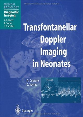
This book examines in detail the role of transfontanellar pulsed and color doppler imaging in the fetus and neonate. After an introductory chapter its use in the normal neonate is considered. Results of the hemodynamic evaluation of 491 newborns aged from 32 weeks of gestation to 9 months by means of pulsed and color doppler are reported.
Venous ultrasound provides pictures of the veins throughout the body. A doppler ultrasound study may be part of a venous ultrasound examination. Doppler ultrasound is a special ultrasound technique that evaluates movement of materials in the body. It allows the doctor to see and evaluate blood flow through arteries and veins in the body.
Subsequent chapters examine the use of transfontanellar doppler imaging in a variety of commonly encountered pathological conditions, including intracranial hemorrhage, hydrocephalus, hypoxic-ischemic brain damage, pericerebral collections, sinus thrombosis, intacranial infections and brain malformations.
The revolution in the imaging of vascular physiology, the diagnosis, and the prognostic evaluation of vascular disease are not based on mor advanced cranial ultrasound: transfontanellar doppler imaging in neonates.
Transfontanellar pulsed and color doppler imaging in the fetus and neonates. After an introductory chapter on the technical aspects of doppler ultrasonography�.
Buy transfontanellar doppler imaging in neonates (medical radiology): read kindle store reviews - amazon.
Transfontanellar doppler imaging in neonates, by a couture and c veyrac, series edited by al baert, k sartor and je youker; 2001. This book was written by pioneers in the field, and contains substantial information from their own outstanding experience, in both series and anecdotes, as well as an extensive literature.
Unlike 1d doppler imaging, which can only provide one-dimensional velocity and has dependency on the beam to flow angle, 2d velocity estimation using doppler ultrasound is able to generate velocity vectors with axial and lateral velocity components. 2d velocity is useful even if complex flow conditions such as stenosis and bifurcation exist.
Ultrasound imaging uses high frequency sound waves to make detailed pictures of internal organs and tissues in real time. There are many different applications of doppler ultrasound imaging, including abdominal, pelvic, musculoskeletal, internal organs (such as the scrotum and prostate) and vascular.
Doppler ultrasound is a special type of ultrasound which is used to look at blood flow. A doppler ultrasound machine has a hand-held scanner which is connected to a computer. It uses soundwaves to make pictures of the blood flow in your major arteries and veins.
We know that many cerebral lesions are of circulatory origin and it is now important to study cerebral hemodynamics by pulsed and color doppler ultrasonography. The revolution in the imaging of vascular physiology, the diagnosis, and the prognostic evaluation of vascular disease are not based on morphological sonographic studies but on the doppler techniques that can display cerebral vessels.
Doppler ultrasound may be used to check blood flow to an area. It may also be needed to check for injuries to blood vessels or assess treatment. Common reasons for use include: look for cause of blood pooling in legs such as valve problems in your leg veins; heart valve defects and congenital heart disease blockage in artery� peripheral artery.
Transfontanellar doppler imaging was until recently an emerging new diagnostic modality that was not widely used in clinical practice. Veyrac have pioneered and incessantly developed the tech and neonatal brain scanning until it has now been matured into an nique of prenatal indispensable tool for the correct management of infants.
In this study, we report normal reference ranges for doppler parameters obtained in a large group of healthy volunteers. Echocardiographic data were acquired using state-of-the-art cardiac ultrasound equipment following doppler acquisition and measurement protocols approved by the european association of cardiovascular imaging.
Doppler imaging takes advantage of the fact that blood moves relative to the direction of acoustic wave propagation to create images based on the strength and direction of the doppler shift in the backscattered signal. Related terms: magnetic resonance imaging; atrial natriuretic peptide.
Transcranial doppler, or transcranial doppler (tcd), uses a sonographic imaging from the of the right middle cerebral artery in the head. The sensitivity of tcd varies from 68 % to 89 % relative to contrast tee, and its specificity from 92 % to 100 % when studying stroke populations.
This book examines in detail the role of transfontanellar pulsed and color doppler imaging in the fetus and neonates. After an introductory chapter on the technical aspects of doppler ultrasonography, its use in the normal neonate is considered. Results of the hemodynamic evaluation of 491 newborns aged from 32 weeks of gestation to 9 months by means of pulsed and color doppler are reported.
Book review transfontanellar doppler imaging in neonates m furness department of organ imaging women's and children's hospital north adelaide, south australia australia.
Cranial ultrasonography and transfontanellar doppler in premature neonates (24–32 weeks of gestation): dynamic evolution and association with a severe adverse neurological outcome at hospital discharge in the aquitaine cohort, 2003–2005.
Clinical doppler ultrasound, 3rd edition expert consult: online and print. Transfontanellar doppler imaging in neonates (medical radiology / diagnostic imaging).
What is a transfontanellar ultrasound? it is an exam used to diagnose brain lesions and predict changes in the newborn's neurological development.
Subsequent chapters examine the use of transfontanellar doppler imaging in a variety of commonly encountered pathological conditions, including intracranial hemorrhage, hydrocephalus, hypoxic-ischemic brain damage, pericerebral collections, sinus thrombosis, intacranial infections, and brain malformations.

Post Your Comments: