Read Advanced Microscopy in Mycology (Fungal Biology) - Tanya Dahms file in ePub
Related searches:
The mycology reference laboratory (mrl) is situated at the phe south west laboratory in bristol. The laboratory provides a comprehensive service for the diagnosis and management of fungal.
Roberson confocal microscopy is an advanced fluorescence microscopy technique that utilizes laser illumination, optical devices, pinholes, computers, and cameras/detectors to perform cell.
Fungal tests are used to help detect and diagnose a fungal infection, to help guide treatment, and/or sometimes to monitor the effectiveness of treatment. For many superficial skin and yeast infections, a clinical examination of the patient and microscopic examination of the sample may be sufficient to determine that a fungal infection is present.
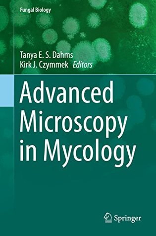
This volume provides insight into the principles underscoring various advanced microscopy methods and how they have been, or have the potential to be, applied to mycology. Offering a comprehensive overview of the confocal principle, confocal laser scanning microscopy and its application to fungal biology, this text also examines the newer.
Skin, hair and nail tissue are collected for microscopy and culture (mycology) t o establish or confirm the diagnosis of a fungal infection. Exposing the site to long-wavelength ultraviolet radiation (wood lamp) can help identify some fungal infections of hair (tinea capitis) because the infected hair fluoresces green.
Skin, hair and nail tissue are collected for microscopy and culture (mycology) to establish or confirm the diagnosis of a fungal infection.
Module 2 - how to use basic microscopy methods on wet mounted samples from a wide diversity of human tissues. Module 3 - an introduction to histology and identification of fungal elements in many human tissues. Module 4 - advanced skills in histology for identification of uncommon and rare fungal diseases.
Oct 4, 2016 only about 20 to 25 species of fungi are common causes of infection. Fungal tests detect infections and sometimes identify the fungus and help.
Quick-freeze, deep-etch electron microscopy reveals the characteristic architecture of the fission yeast spore.
Mycology in a developing country, it provides clear guidance in a very readable form. It will hopefully contribute toward enhancing the detection of fungal diseases and increasing the cadre of enthusiastic medical mycologists across the globe. Rumina hasan abdulaziz hussainali shariff professor mbbs, phd, frcpath.
Nonlinear microscopy as a tool to study growth and development of fungi.
When unable to observe the myco-microcosms my curiosity hit a bit of a roadblock. Being a visual learner, i wanted to witness every stage in a fungus's life cycle.
Microscopy, however, cannot determine the specific cause of infection. While gram stain lacks sensitivity, fluorescent brighteners (calcofluor white, or blankophor), which bind to chitin in the fungal cell wall, are a rapid means of scanning samples for fungal hyphae, and enhance morphology assessment (16).
These studies greatly increased our knowledge of the fungal biota in peanuts and pecans, two major crops in georgia. In addition, to strengthen the mycology curriculum at the main campus in athens, he commuted twice weekly one quarter per year to teach an advanced mycology course, since there was no mycologist in athens at the time.
The aim of advanced microscopy in mycology is to describe the latest advances in microscopic methods, including integrated techniques, as applied to mycology.
Jun 24, 2020 based on deep neural networks and fisher vector (advanced bag-of-words method) to classify microscopic images of various fungi species.
Fungal infections (also called mycoses) represent the invasion of tissues by one or more species of fungi. They range from superficial, localized skin conditions to deeper tissue infections to serious lung, blood or systemic diseases.
At the tip of each fungal hypha lies a region of active growth. Using advanced, super-resolution microscopy techniques to visualise the activities of living cells.
Medical mycology laboratories are most familiar with the asexual reproduction stage. However, sexual structures are the baseline for fungal taxonomy and nomenclature. 72 during infections, most pathogenic moulds show only hyphal elements or other, mostly non-specific, structures in host tissue.
The source is frequently the gastrointestinal tract or indwelling catheters particular with hyperalimentation. 18 a blood fungus culture is useful to define invasive disease; however, proof of invasive candida infection requires direct cystoscopic or operative visualization, fungus balls, pyelonephritis, or histological evidence of mucosa.
The field of mycology is poised to exploit the many recent advances in microscopic tools and instrumentation for cell biology. This chapter first outlines the latest developments in biosensors that.
Microscopy: potassium hydroxide (koh) stain is a commonly-used method because it is inexpensive and easy to perform. Nail clippings or scrapings are placed in a drop of koh and examined under a microscope for the presence of fungal elements.
Fundamentals of mycology including ecology, biology and classification of fungi will be discussed. Surface samples such as tapes, bulks and swabs are analyzed using dme techniques for variety of fungal genera in indoor environments. Participants also review laboratory reports and learn interpretation of fungal data.
Jul 25, 2017 direct microscopic examination of fungi in clinical specimens relies on both bright -field and phase-contrast microscopy, as well as multiple.
The aim of advanced microscopy in mycology is to describe the latest advances in microscopic methods, including integrated techniques, as applied to mycology. Each chapter will provide a brief overview of a particular microscopic method with associated advantages and limitations, the research questions that can be appropriately addressed using.
Many of the fungal infections are caused by opportunistic fungi, meaning that they only cause infection if the conditions are just right. Mycology is the department of microbiology that studies and identifies fungi, including those that are pathogenic to humans and are possible causes of disease.
Dec 12, 2014 the fungal kingdom can be an inspiration for even more. Highly advanced mycological research has generated knowledge, conceptual polarisome meets spitzenkörper: microscopy genetics, and genomics converge.
The field of mycology is poised to exploit the many recent advances in microscopic tools and instrumentation for cell biology. This chapter first outlines the latest developments in biosensors that will prove useful for targeting specific events in the context of single fungal cells, mycelia, or fungal-plant systems.
Donald danforth plant science center; fungal partners secrete bioactive molecules such as small peptide effectors.
This website contains information on the identification and management of human and animal fungal infections. The site provides a range of educational materials including a mould identification self assessment module, descriptions of fungal pathogens and diseases, antifungal susceptibility data and links to societies and to other mycology sites.
Skin scrapings suspected to contain dermatophytes or pus from a lesion can be mounted in koh on a slide and examined directly under the microscope. Serology may be helpful when it is applied to a specific fungal disease. Direct fluorescent microscopy may be used for identification.
Digital microscopy combined with the rapid proliferation of image sharing platforms offers a unique opportunity to harness this shared visual content for expanding education about key fungal spore type morphologies of interest to the environmental mycology community.
April 6, 2019 acharya tankeshwar lab diagnosis of fungal disease, mycology 4 early diagnosis of fungal infection is critical for effective treatment. For many decades, culture, direct microscopy, and histopathology have been the foundation for the diagnosis of fungal infections.
Electron microscopy is used to observe the ultra-structure and fungal pathogens for species identification and study of pathogenesis. For translational medical mycology, molecular biology and individualized treatment should include both the host and the fungus.
Mar 7, 2013 proliferation of fungal hyphae was primarily through pits. In petioles with more advanced stages of necrosis, hyphae were also found in the pith.
Molecular mycology: current approaches to fungal pathogenesis august 14 – august 29 microscopy.
Buy advanced microscopy in mycology (fungal biology) on amazon.
Charles darwin house, 12 roger street, london, wc1n 2ju email: admin@britmycolsoc.
Advanced microscopy in mycology part of the fungal biology book series ( fungbio) confocal laser-scanning microscopy in filamentous fungi.
2017-11-01 [pdf] advanced microscopy in mycology (fungal biology) - removed; 2020-02-26 management of fungal pathogens in pulses: current status and future challenges (fungal biology) 2020-02-23 recent developments on genus chaetomium (fungal biology) 2019-12-25 biology of macrofungi (fungal biology).
Isolation and identification (additional charges/cpt code[s] may apply) if culture results warrant.
Direct microscopy is a rapid method for distinguishing many fungal infections using microscopic methods with specific stains. This is the first online microscopy course for fungal infections. Direct microscopy is a rapid method for distinguishing many fungal infections using microscopic methods with specific stains. This is the first online microscopy course for fungal infections.
Dimensional (3d) electron microscopy and fluorescent biosensors. We consider cium ion dynamics using organic fluorescent dyes in fungi has met with limited.
includes 60 images of fungi collected using a variety of techniques provides the most comprehensive and up-to-date review of state of the art microscopy.
Dec 6, 2012 laboratory protocols in fungal biology is a valuable tool for both beginner research workers and dahms / advanced microscopy in mycology.
Confocal microscopy is a revolution- ary advance in light microscopy. Utilizing the latest laser, computer, and imaging technologies, the technique offers biologists a unique form of cell and/or subcellular visualization.
5— fering from cancer or advanced diabetes, and patients undergoing the hyphae are easily seen by light microscopy, and are termed clamp.
Caused by the fungus histoplasma, which lives in the environment, often in association with large amounts of bird or bat droppings. Fungal diseases that affect people with weakened immune systems weakened immune systems can’t fight off infections as well, due to conditions such as hiv, cancer, organ transplants, or certain medications.
Using a combination of environmental dna sequencing and fluorescence microscopy, a new component of the fungal tree of life was identified and this wider.
Introduction mycology - the study of fungi fungi - molds and yeasts molds - exhibit filamentous type of growth yeasts - pasty or mucoid form of fungal growth 50,000 + valid species; some have more than one name due to minor.
All sections can of course be mounted without any staining, and this can be in distilled water, however, potassium hydroxide (k0h), or ammonium hydroxide is generally preferred. Potassium hydroxide does not keep as well as ammonium hydroxide as the former will crystallise out in time.
Confocal microscopy is a revolutionary advance in light microscopy. It is particularly advantageous for examining thick fungal samples or in host- pathogen.
Oct 12, 2020 confocal microscopy has been successfully applied to answer a wide variety of fungal biology questions including cell and cytoplasmic dynamics.
Dec 1, 2017 the identification and nomenclature of fungi is quite difficult. The nomenclature and classification of fungal species are usually based on their.
This lecture covers the basics of microscopy to identify unknown fungi samples.
The ability of fungi to invade plant and animal tissue was observed in early 19th century but the first documented animal infection by any fungus was made by bassi, who in 1835 studied the muscardine disease of silkworm and proved the that the infection was caused by a fungus beauveria bassiana.
Histoplasma capsulatum is an intracellular, thermally dimorphic fungi (grows as a yeast in body temperature/37°c in humans, mammals or in culture media and as mold in 25°c in environment/culture media) of medical importance that can survive within macrophages for an extended period. This fungal pathogen is associated with birds or bat droppings.
The field of diagnostic mycology represents much more than culture and microscopy and is rapidly embracing novel techniques and strategies to help overcome the limitations of conventional approaches. Commercial molecular assays increase the applicability of pcr testing and may identify markers of antifungal resistance, which are of great.
Numerous images enhance the descriptions of identifying characteristics by illustrating the appearance of fungal colonies on media and microscopic appearance.
Start studying (mycology) common fungal oppurtunists microscopy. Learn vocabulary, terms, and more with flashcards, games, and other study tools.
Light microscopy is a necessary skill for accurate fungal identification of many species. The aim of this section is to gather some notes and articles to help field mycologists increase their knowledge on the subject. The first page has been submitted by peter smith and covers the range of chemical reagents used in fungal.
The mycology reference laboratory (mrl) provides a comprehensive laboratory service for the diagnosis and management of fungal infections.
Apr 3, 2020 “super-resolution microscopy: sim, sted and localization microscopy,” in advanced microscopy in mycology fungal biology, eds dahms.
Director of the center for advanced microscopy and imaging; instructor microscopy, ultrastructural imaging, mycology, and developmental morphology ultrastructural reproductive development of members of the fungal class zygomycete.
The course is designed to teach the identification of fungal infection by direct (wet) module 4 covers advanced skills in histology for identification of uncom.
Fungal tests are used to help detect and diagnose a fungal infection, to help guide treatment, and/or to monitor the effectiveness of treatment. For many superficial skin and yeast infections, a clinical examination of the affected body part(s) and microscopic examination of the sample may be sufficient to determine that a fungal infection is present.
Mycology: characteristics of fungi, fungal diseases, and more see online here almost every clinically active doctor is confronted with fungal infections during their working life. It can be foot or nail fungi if they are a family doctor, fungal pneumonia if they are a haemato-oncologist, or irritant diaper dermatitis if they are a pediatrician.
8 future directions in advanced mycological microscopy 145 genetic encoding can help circumvent the recalcitrant properties of the fungal cell and its tendency to sequester or otherwise compartmentalize/degrade exog-.
In the mycology laboratory, the material is examined using potassium hydroxide (koh) to dissolve keratinocytes, then staining the preparation with blue or black ink to identify koh-resistant mycelium and arthrospores by direct microscopy. Fungal elements are sometimes difficult to find, especially if the tissue is very inflamed, so a negative.
Medical mycology clinical lab courses are also limited in this specialty but we expected that to be changing with complementary fungal dna research and analysis. But we were later in for a surprise update� rec ent study (dec 2003-abstract) reports less training now than before in medical mycology - steinbach.
Offering a comprehensive overview of the confocal principle, confocal laser scanning microscopy and its application to fungal biology, this text also examines the newer sophisticated fluorescence-based methods of technology.
Mushrooms, also known as fungi or toadstools, are the spore-bearing fruiting body of a fungus. The mushroom typically grows above ground or on top of its food.
Post Your Comments: